For years, scientists have understood the necessity of a complex network of blood vessels for nourishing the retina cells, crucial for our vision. The process of creating such an intricate structure, however, remained a mystery until researchers at UC San Francisco made a remarkable discovery. They found a unique kind of neuron that governs the formation of this lattice structure, as reported in the May 23, 2024 issue of Cell.
This groundbreaking discovery could pave the way for innovative therapies for diseases associated with poor blood circulation in the eyes and brain. Dr. Xin Duan, an associate professor of ophthalmology and the senior author of the study, stated: “Being able to witness retinal neurons guiding blood vessels directly to form these intricate 3-D lattices is a first. This knowledge brings us a step closer to the potential of repairing or redirecting them when necessary.”
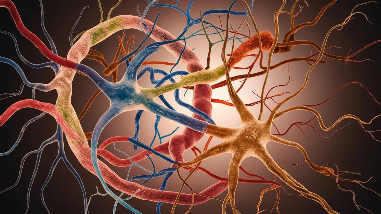
In their research, the team utilized newborn mice, whose eyes still require a few weeks for complete development. Kenichi Toma, a fellow researcher, marked the retinal neurons closest to the blood vessels with a specially designed protein that emits a green glow under ultraviolet light. This allowed the team to monitor the formation of the lattice structure.
They identified a unique subset of neurons, referred to as perivascular neurons. These neurons surround and guide the growing blood vessels, orchestrating them to form the lattice. These perivascular neurons produce a protein, PIEZO2, that allows them to sense when they are in contact with another cell.
When these perivascular neurons in mice could not produce PIEZO2, they could not maintain contact with the blood vessels, resulting in a disorganized, tangled growth that hindered blood flow. As a result, the surrounding nerve cells degraded due to oxygen deprivation, making the mice more susceptible to stroke-like injuries.
Interestingly, Duan discovered that these neurons play a similar role in forming a network of blood vessels in the cerebellum, a brain region responsible for coordination, language, and sensory perception. This repetitive pattern in the brain suggests that damage to this lattice could contribute to various neurodegenerative diseases.
By collaborating with developmental biologist Arnold Kriegstein, the team confirmed the existence of perivascular retinal neurons in humans as well.
The researchers used a cutting-edge technique called multiphoton microscopy to create 3-D images of retinal blood networks without disturbing the eye. This technique aided Toma in creating rotating movies that captured the lattice from every angle, demonstrating its breakdown in the absence of PIEZO2.
This breakthrough discovery could lead to new treatments for neurodegenerative diseases by ensuring that neurons, which require high energy, maintain a healthy blood supply. “While many are trying to understand how to grow neurons, the challenge lies in figuring out how to form the intricate networks of blood vessels necessary to support them. That’s the question we’re attempting to answer,” Duan concluded.
Comments are closed for this post.