The nucleus of a cell, well-known for its role in genetic regulation, has been found to play a crucial role in tissue and organ formation during embryonic development. This discovery was made by a research group led by biophysicist Otger Campàs at both UC Santa Barbara and the Physics of Life Excellence Cluster of TU Dresden. This revelation brings a new dimension to our understanding of the cell’s nucleus, expanding its function beyond genetic regulation to include tissue structuring.
Previous thought suggested that tissue stiffness was determined by surface interactions between cells, not by internal components like organelles. However, Campàs and his team observed that the stiffness of zebrafish retina tissue was significantly influenced by the arrangement of cell nuclei. This unexpected finding, subsequently published in Nature Materials, opens a new line of inquiry into how cells drive embryonic development.
Inside every cell, various structures called organelles undertake essential functions. Similar to the factories and roads in a city, these countless organelles work within cells to ensure their proper operation. Traditionally, due to them being contained within cells, organelles were not thought to be directly involved in the construction of organs during embryonic development. However, the recent findings challenge this belief.
The cell’s nucleus, an organelle renowned for its role in controlling cellular activity, is also the largest and stiffest structure within the cell. Hence, Campàs hypothesized that the nucleus could influence the physical structure of the tissue alongside processing information.
Campàs and his team previously discovered that groups of cells behave like foam during development, which can either be ‘jammed’ to fix the tissue structure and establish its shape, or ‘melted’ to allow tissue to flow and form shapes. In their recent research, they found that a transition from solid to fluid could be governed by the relative sizes of the nucleus and the cell.
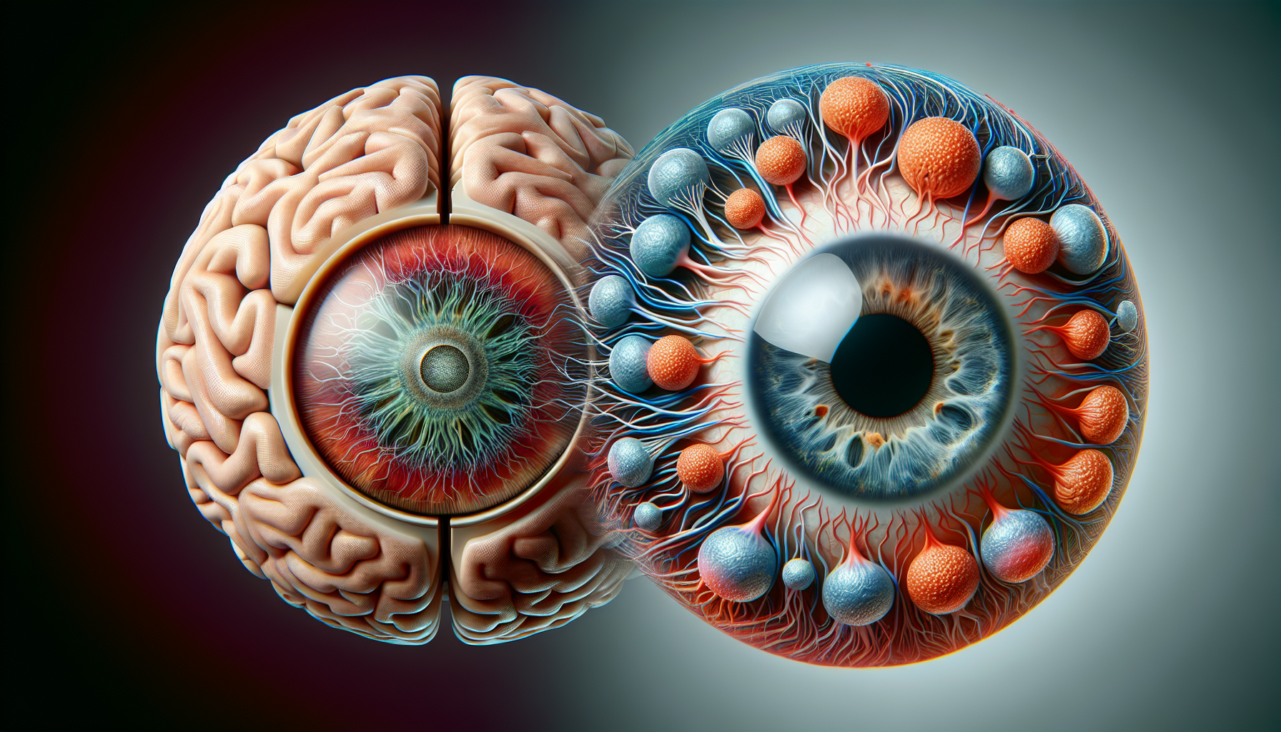
The researchers found that the stiffness of eye and brain tissues was directly regulated by the nucleus when it occupied a significant portion of the cell space. They also observed that when the nuclei were packed tightly, they arranged the cells into nearly perfect arrays. This discovery indicates that the nucleus, and not the cell surface, determines both tissue mechanics and cellular ordering. This challenges current understanding and highlights a novel role for the nucleus in controlling tissue organization and mechanics.
To delve deeper into this phenomenon, the research team utilized zebrafish as their subject of study. These creatures are transparent during their embryonic stages and mature rapidly, making them an excellent model for studying organ formation in 3D. The researchers measured structural features and quantified cell movements, particularly focusing on the developing retina and brain of zebrafish.
The team found that changes in cell and nuclear sizes during crucial developmental stages caused the nuclei to become ‘jammed’ in place, tightly packed by surrounding cells. This tight packing resembled the arrangement of coffee beans in a jar and seemed to be essential for the eye’s function. They also discovered that the brain tissues exhibited a similar ‘nuclear jamming’, indicating a wider role for the nucleus in shaping the architecture of neural tissues.
This pioneering work not only unveils a new role for the nucleus in tissue organization but also suggests that defects at the nuclear level could lead to diseases linked with impaired tissue architecture. Such insights bring us a step closer to fully understanding how cells build organs during embryonic development.
Comments are closed for this post.